
Proteins and protein complexes can be thought of as microscopic machines at work inside our cells. To function properly, they must adopt the right shape by their correct folding. Disruption of this process can lead to disease. The challenge is that these structural changes happen very fast and on an extremely small scale – fractions of a nanometer, or less than a billionth of a meter – making them very difficult to observe directly.
How do you see something that can’t be seen?
The same protein can behave differently depending on its environment – for example, the pH level, salt concentration, or the presence of other molecules.
In some conditions, it will be more tightly folded and stable; in others, it will more easily change its structure. This, in turn, affects how it interacts with other proteins, drugs, or cells.
Since the late 1990s, scientists have been able to observe individual protein molecules as they unfold and refold. The University of Warsaw team, however, aims to go further and capture the subtle intermediate states that occur during these changes.
“Our approach is not to directly ‘see’ the protein structure, but to infer it from its mechanical properties. We measure local stiffness and what we call internal friction within the molecule. This refers to how quickly energy is dissipated by a given chemical structure. As the structure changes, these parameters change as well, allowing us to infer its state – including intermediate states during the folding process,” explains Prof. Robert Szoszkiewicz from the Laboratory of Physicochemistry of Materials at the University of Warsaw Biological and Chemical Research Centre.
Every structural change, even a very small one, affects how easily a protein can be stretched and how much energy it loses as it moves. It is a bit like a musical string: we cannot see inside it, but we can tell how tightly it is tuned from the sound it makes.
A closer look at a single molecule
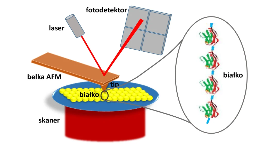
A key tool in this research is the atomic force microscope (AFM). It works much like a vinyl record player, except that instead of reading sound from a vinyl record, it probes the properties of individual molecules. In an AFM, an extremely small needle – ending in a tip just a few atoms wide – can be in contact with the sample and respond to its properties. In a standard contact-mode, this allows researchers to image surfaces, but in protein studies a different mode is used, known as force spectroscopy. Here, protein molecules are attached to a surface, and the microscope tip can grab their other end and gradually stretch them.
“We can, for example, apply a physiologically relevant force (one that proteins respond to – editor’s note) of around 100 piconewtons (a piconewton is one trillionth of a newton, the unit of force – editor’s note). We then observe how the protein unfolds, and gradually reduce the force to follow how it refolds. The most interesting part happens ‘along the way,’ when we track changes in mechanical parameters along a refolding trajectory,” explains Prof. Szoszkiewicz.
To ensure they are truly observing the behavior of a specific protein, researchers often covalently link many identical copies of a given protein into a longer chain. This way, the same signals are repeated multiple times during the experiment, making it easier to distinguish genuine measurements from random noise.
“The key point is that we can precisely control very small forces acting on a protein and observe how it mechanically responds to these forces under different physicochemical conditions – for example, in different solutions. This gives us access to information that was previously practically inaccessible,” the scientist adds.
Different spatial configurations of the proteins link to their distinct mechanical properties.
Importantly, the equipment used in these studies was built by the University of Warsaw scientists themselves.
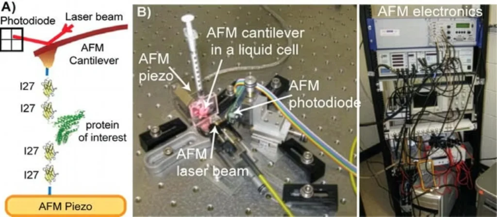
podpis: A modified AFM microscope built in Prof. Szoszkiewicz’s laboratory at the University of Warsaw, used to manipulate individual protein molecules—here shown using the model protein I27_4.
When vibrations reveal more than images can
The most original aspect of the method lies in how these parameters are measured. The AFM tip is not just a “sensor” but behaves like a vibrating beam with specific resonant frequencies – frequencies at which the beam oscillates with the largest amplitudes.
“Every oscillating system has certain frequencies at which it vibrates most easily. We track several of these at the same time and observe how they change during the experiment,” explains Prof. Szoszkiewicz.
When the microscope tip is attached to a protein, even very small changes in its structure affect how the AFM cantilever vibrates. Thus, any shifts in its resonant frequencies become a sensitive indicator of molecular shape changes of the protein molecule. This allows scientists to determine whether a given protein is transitioning into a different spatial arrangement of its components.
“This may seem surprising, because we are dealing with a relatively large element – a microscope cantilever tens of micrometers long – connected to a very small object, a protein only a few nanometers in size. And yet the protein’s effect on the cantilever’s vibrations is measurable,” the scientist notes.
The data obtained in this way do not yet allow the protein structure to be directly “seen,” but they form the basis for further analysis. The next step is to combine the experimental results with computer simulations.
“Different protein configurations have distinct stiffness and internal friction. If we combine our measurements with molecular dynamics simulations, we can begin to infer which configurations are most likely for a given set of measured mechanical parameters,” he explains.
What have scientists observed?
Researchers have developed a model that allows them to extract from microscope measurements how stiff a protein is and how much energy it dissipates during structural changes.
To make this possible, they first had to carefully describe the behavior of the AFM tip itself, which continuously vibrates during experiments, and in relevant environments such as water.
Next, they could start to study the properties of the tip-protein systems. The studies showed that protein properties can change very dynamically under applied force. In particular, when a protein is stretched, it becomes noticeably stiffer and loses more energy as its structure transitions between nearby configurations. One of the key results obtained so far is that the team also managed to capture short-lived intermediate states between the folded and unfolded forms for one particular proteins. Until now, these transient stages have been extremely difficult to observe.
Key insight: The crucial role of the environment
Another of the most interesting findings is that the observed internal friction of the proteins does not arise solely from the protein’s structure itself. It is largely determined by interactions between the protein and its surroundings, especially water. This means that folding is not purely an “internal matter” of the molecule, but is strongly influenced by the environment in which it takes place. From that perspective internal friction can be linked to molecular-level processes, such as changes in hydrogen-bond networks in aqueous environments.
The developed method makes it possible to study proteins in a way that was previously practically inaccessible: at the level of a single molecule, during an ongoing process, and with sensitivity to very subtle structural changes.
A step toward new applications
Although this is basic research focused primarily on understanding how proteins behave, it may in the future also find applications in medicine, particularly in targeted cancer therapies.
Modern cancer treatment strategies increasingly rely on short protein fragments called peptides. They can act like a “key” designed for a specific “lock” – recognizing particular cancer cells and precisely targeting them.
The problem is that the environment inside a tumor is very different from laboratory conditions. As a result, these molecules can change their shape and behave in ways not intended by the scientists who designed them.
“The pH changes, ion concentration varies, and many different molecules are present. As a result, peptides can adopt structures other than those originally designed and stop functioning as expected,” explains Prof. Szoszkiewicz.
One possible solution is to better understand how specific molecules behave in different environments. This is where methods developed at the University of Warsaw may play an important role.
“We can study how the mechanical properties of peptides change under varying conditions and then, combined with computer simulations, try to reconstruct their most likely structures. This can help identify which of them will work best in a given environment,” the scientist explains.
At the same time, the method has its limitations. It works best for small proteins and peptides. In larger structures, different configurations can produce similar values of the measured mechanical parameters, making interpretation more difficult. Another challenge is time resolution: protein folding processes can occur very quickly.
“This means we are not always able to capture all short-lived intermediate states. But from an application perspective, the more stable final structures are often more important – and those we can study,” says Prof. Szoszkiewicz.
Despite these limitations, the approach remains unique and is being developed in only a few research centers worldwide.
“It is a demanding method, requiring time and resources. But it is precisely such approaches that sometimes lead to the most interesting results,” the scientist emphasizes.
What’s next?
The current method makes it possible to observe mechanical changes in proteins with sub-second time resolution. That is significant, but many relevant processes occur much faster – for example, the brief breaking and forming of chemical bonds that help a protein change its shape.
The next step is therefore to improve this temporal resolution and further develop models that will allow researchers to link mechanical properties even more precisely to specific changes in molecular structure. The ultimate goal is to fully reconstruct the folding process step by step at the level of a single molecule.
“This is a project that requires time, a team, and funding. Without sufficient resources, it is difficult to speed it up. In the future, collaboration with medical sciences will also be crucial if the method is to find therapeutic applications,” says Prof. Szoszkiewicz, adding that despite the challenges, such work is worth pursuing: “In science, the most demanding problems often turn out to be the most interesting.”
The text was originally published in Polish on the Serwis Naukowy UW website on May 14, 2026.